 Yuji Ikegaya
open access DATA archive
FUNCTIONAL MULTINEURON CALCIUM IMAGING (fMCI)
fMCI is a functional imaging technique with multicell loading of calcium fluorophores, which was originally introduced by Yuste and Katz (Neuron 6:333-344, 1991). fMCI has unique advantages, including: i) recording en masse from hundreds of neurons in a wide area, ii) single-cell resolution, iii) identifiable location of neurons, and iv) detection of non-active neurons during the observation period. For details, see our method paper.
Methods
Rat slice cultures were loaded with Oregon Green 488 BAPTA-1AM, and calcium signals were optically recorded from the CA3 pyramidal cell layer in aCSF consisting of (in mM): 127 NaCl, 26 NaHCO3, 3.3 KCl, 1.24 KH2PO4, 1.0-2.0 MgSO4, 1.0-2.0 CaCl2 and 10 glucose. Fluorophores were excited at 488 nm and visualized with a 507-nm long-pass emission filter. Images were captured at 10-2000 frames/s using a Nipkow spinning-disk confocal microscope (CSU-X1; Yokogawa Electric, Tokyo, Japan), a cooled CCD camera (iXon DU897; Andor, Belfast, Northern Ireland, UK), an upright microscope (ECLIPSE FN1, Nikon, Tokyo, Japan), and a water-immersion objective (16X, 0.80 NA, CFI75LWD16xW, Nikon).
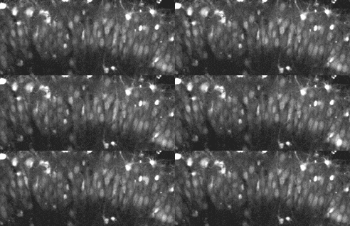
Fig. 1 Confocal movie taken from the CA3 pyramidal cell layer of a hippocampal
slice incubated in Oregon Green 488 BAPTA-1AM.
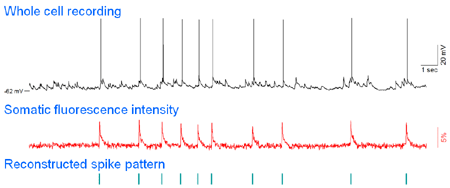
Fig. 2 Simultaneous whole-cell patch-clamp recording and calcium imaging. Action
potentials (top), but not synaptc inputs, are reliably reflected in somatic
calcium transients (middle), and thus, the timings of spikes can be reconstructed
from the onsets of calcium transients without electrophysiologic recordings
(bottom).
Data Representation
Data are given in the TEXT file format. A part of DATA_001.txt is shown below.
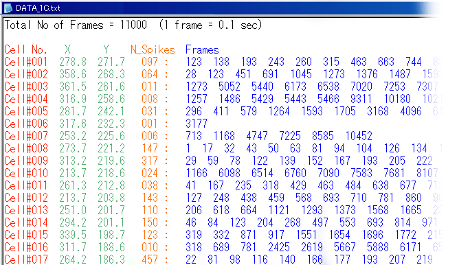
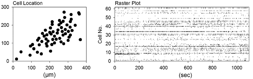
In this data file, the movie length is 18 min 20 sec (=11000 frames at 0.1 sec per frame). Each horizontal row in the Data part represents the data of a single cell. This file contains 62 cells (rows). The first ' red' column indicates the cell number (assigned arbitrarily). The next two 'green' columns indicate the 'X versus Y' coordinate of the cell location (unit: micrometer). The 'orange' number indicates the number of the total activity of the cell during the movie. For example, Cell1 showed 97 activities during the recording period of 18 min 20 sec. If this value is zero, the neuron was silent. The following 'blue' columns indicate individual activity times of the cell, i.e., frames at which the neuron fired. For example, Cell1 was activated at frames 123, 138, 193, 260, ..., that is, it fired at 12.3, 13.8, 19.3, 26.0, ..., sec after the beginning of the movie.
As a result, the cell map and the activity rastergram can be reconstructed from DATA_001.txt as follows.
Archive (for download, right-click on the filename)
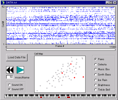
To confirm whether you correctly reconstructed spike trains from these data, the Visual Basic program Raster_Reconstractor.exe (76 kb) may be useful (see below). Because this program is written in Visual Basic 6.0 for Microsoft Windows PCs, VB6 runtime is required to run it. The runtime is distributed at Microsoft Download Center (free).
References
Ikegaya, Y., Aaron, G., Cossart, R., Aronov, D., Lampl, I., Ferster, D., and Yuste, R. Synfire chains and cortical songs: Temporal modules of cortical activity. Science, 304:559-564, 2004.
Sasaki, T., Matsuki, N., Ikegaya, Y. Metastability of active CA3 networks. J. Neurosci., 27:517-528, 2007.
Takahashi, N., Sasaki, T., Matsumoto, W., Matsuki, N. and Ikegaya, Y. Circuit topology for synchronizing neurons in spontaneously active networks.Proc. Natl. Acad. Sci. U. S. A., 107:10244-10249, 2010.
Review
Takahashi, N., Sasaki, T., Usami, A., Matsuki, N., Ikegaya, Y. Watching neuronal circuit dynamics through functional multineuron calcium imaging (fMCI). Neurosci. Res., 58:219-225, 2007. (PDF)
Protocol
Takahashi, N., Oba, S., Yukinawa, N., Ujita, S., Mizunuma, M., Matsuki, N., Ishii, S. and Ikegaya, Y. High-speed multineuron calcium imaging using Nipkow-type confocal microscopy. Curr. Protoc. Neurosci., 2:Unit2.14, 2011.
(PDF)
Note
Although these files are freely available for analysis, we still own the copyright of the original (not analyzed) data and may claim coauthorship when the analyzed data are published elsewhere. The contact informasion is:
Yuji IKEGAYA
Laboratory of Chemical Pharmacology,
Graduate School of Pharmaceutical Sciences,
The University of Tokyo.
7-3-1 Hongo, Bunkyo-ku, Tokyo 113-0033, Japan.
TEL: 03-5841-4780, FAX: 03-5841-4786
yuji@ikegaya.jp
FUNCTIONAL MULTINEURON CALCIUM IMAGING (fMCI)
fMCI is a functional imaging technique with multicell loading of calcium fluorophores, which was originally introduced by Yuste and Katz (Neuron 6:333-344, 1991). fMCI has unique advantages, including: i) recording en masse from hundreds of neurons in a wide area, ii) single-cell resolution, iii) identifiable location of neurons, and iv) detection of non-active neurons during the observation period. For details, see our method paper.
Methods
Rat slice cultures were loaded with Oregon Green 488 BAPTA-1AM, and calcium signals were optically recorded from the CA3 pyramidal cell layer in aCSF consisting of (in mM): 127 NaCl, 26 NaHCO3, 3.3 KCl, 1.24 KH2PO4, 1.0-2.0 MgSO4, 1.0-2.0 CaCl2 and 10 glucose. Fluorophores were excited at 488 nm and visualized with a 507-nm long-pass emission filter. Images were captured at 10-2000 frames/s using a Nipkow spinning-disk confocal microscope (CSU-X1; Yokogawa Electric, Tokyo, Japan), a cooled CCD camera (iXon DU897; Andor, Belfast, Northern Ireland, UK), an upright microscope (ECLIPSE FN1, Nikon, Tokyo, Japan), and a water-immersion objective (16X, 0.80 NA, CFI75LWD16xW, Nikon).
|

 |
Data Representation
Data are given in the TEXT file format. A part of DATA_001.txt is shown below.
In this data file, the movie length is 18 min 20 sec (=11000 frames at 0.1 sec per frame). Each horizontal row in the Data part represents the data of a single cell. This file contains 62 cells (rows). The first ' red' column indicates the cell number (assigned arbitrarily). The next two 'green' columns indicate the 'X versus Y' coordinate of the cell location (unit: micrometer). The 'orange' number indicates the number of the total activity of the cell during the movie. For example, Cell1 showed 97 activities during the recording period of 18 min 20 sec. If this value is zero, the neuron was silent. The following 'blue' columns indicate individual activity times of the cell, i.e., frames at which the neuron fired. For example, Cell1 was activated at frames 123, 138, 193, 260, ..., that is, it fired at 12.3, 13.8, 19.3, 26.0, ..., sec after the beginning of the movie.
As a result, the cell map and the activity rastergram can be reconstructed from DATA_001.txt as follows.
Archive (for download, right-click on the filename)
| data # | movie length | frame rate | number of neurons | region | temperature | |
|---|---|---|---|---|---|---|
| DATA_001.txt | 1100 sec | 10 Hz | 62 | hippocampal CA3 | 32 ºC | |
| DATA_002.txt | 700 sec | 10 Hz | 126 | hippocampal CA3 | 32 ºC | |
| DATA_003.txt | 600 sec | 10 Hz | 226 | hippocampal CA3 | 32 ºC | |
| DATA_004.txt | 1200 sec | 10 Hz | 64 | hippocampal CA3 | 32 ºC | |
| DATA_005.txt | 610 sec | 10 Hz | 88 | hippocampal CA3 | 32 ºC | |
| DATA_006.txt | 1200 sec | 10 Hz | 95 | hippocampal CA3 | 32 ºC | |
| DATA_007.txt | 310 sec | 10 Hz | 68 | hippocampal CA3 | 32 ºC | |
| DATA_008.txt | 1200 sec | 10 Hz | 93 | hippocampal CA3 | 32 ºC | |
temperature dependence |
||||||
| DATA_009.txt | 180 sec | 100 Hz | 169 | hippocampal CA3 | 24 ºC | |
| DATA_010.txt | 180 sec | 100 Hz | 106 | hippocampal CA3 | 24 ºC | |
| DATA_011.txt | 180 sec | 100 Hz | 135 | hippocampal CA3 | 24 ºC | |
| DATA_012.txt | 180 sec | 100 Hz | 121 | hippocampal CA3 | 24 ºC | |
| DATA_013.txt | 180 sec | 100 Hz | 120 | hippocampal CA3 | 24 ºC | |
| DATA_014.txt | 180 sec | 100 Hz | 156 | hippocampal CA3 | 28 ºC | |
| DATA_015.txt | 180 sec | 100 Hz | 105 | hippocampal CA3 | 28 ºC | |
| DATA_016.txt | 180 sec | 100 Hz | 134 | hippocampal CA3 | 28 ºC | |
| DATA_017.txt | 180 sec | 100 Hz | 112 | hippocampal CA3 | 28 ºC | |
| DATA_018.txt | 180 sec | 100 Hz | 134 | hippocampal CA3 | 28 ºC | |
| DATA_019.txt | 180 sec | 100 Hz | 127 | hippocampal CA3 | 32 ºC | |
| DATA_020.txt | 180 sec | 100 Hz | 179 | hippocampal CA3 | 32 ºC | |
| DATA_021.txt | 180 sec | 100 Hz | 150 | hippocampal CA3 | 32 ºC | |
| DATA_022.txt | 180 sec | 100 Hz | 125 | hippocampal CA3 | 32 ºC | |
| DATA_023.txt | 180 sec | 100 Hz | 135 | hippocampal CA3 | 32 ºC | |
| DATA_024.txt | 180 sec | 100 Hz | 105 | hippocampal CA3 | 36 ºC | |
| DATA_025.txt | 180 sec | 100 Hz | 157 | hippocampal CA3 | 36 ºC | |
| DATA_026.txt | 180 sec | 100 Hz | 137 | hippocampal CA3 | 36 ºC | |
| DATA_027.txt | 180 sec | 100 Hz | 120 | hippocampal CA3 | 36 ºC | |
| DATA_028.txt | 180 sec | 100 Hz | 152 | hippocampal CA3 | 36 ºC | |
| DATA_029.txt | 180 sec | 100 Hz | 129 | hippocampal CA3 | 40 ºC | |
| DATA_030.txt | 180 sec | 100 Hz | 106 | hippocampal CA3 | 40 ºC | |
| DATA_031.txt | 180 sec | 100 Hz | 187 | hippocampal CA3 | 40 ºC | |
| DATA_032.txt | 180 sec | 100 Hz | 160 | hippocampal CA3 | 40 ºC | |
| DATA_033.txt | 180 sec | 100 Hz | 161 | hippocampal CA3 | 40 ºC | |
| DATA_034.txt | 180 sec | 200 Hz | 74 | hippocampal CA3 | 32 ºC | |
fast scanning |
||||||
| DATA_035.txt | 130 sec | 500 Hz | 60 | hippocampal CA3 | 32 ºC | |
| DATA_036.txt | 130 sec | 500 Hz | 88 | hippocampal CA3 | 32 ºC | |
| DATA_037.txt | 130 sec | 500 Hz | 88 | hippocampal CA3 | 32 ºC | |
| DATA_038.txt | 130 sec | 500 Hz | 64 | hippocampal CA3 | 32 ºC | |
| DATA_039.txt | 130 sec | 500 Hz | 96 | hippocampal CA3 | 32 ºC | |
| DATA_040.txt | 130 sec | 500 Hz | 137 | hippocampal CA3 | 32 ºC | |
| DATA_041.txt | 130 sec | 500 Hz | 87 | hippocampal CA3 | 32 ºC | |
| DATA_042.txt | 130 sec | 500 Hz | 95 | hippocampal CA3 | 32 ºC | |
| DATA_043.txt | 130 sec | 500 Hz | 85 | hippocampal CA3 | 32 ºC | |
| DATA_044.txt | 130 sec | 500 Hz | 131 | hippocampal CA3 | 32 ºC | |
| DATA_045.txt | 130 sec | 500 Hz | 77 | hippocampal CA3 | 32 ºC | |
| DATA_046.txt | 130 sec | 500 Hz | 53 | hippocampal CA3 | 32 ºC | |
| DATA_047.txt | 130 sec | 500 Hz | 72 | hippocampal CA3 | 32 ºC | |
| DATA_048.txt | 130 sec | 500 Hz | 60 | hippocampal CA3 | 32 ºC | |
long-time movie |
||||||
| DATA_049.txt | 3300 sec | 10 Hz | 140 | hippocampal CA3 | 32 ºC | |
To confirm whether you correctly reconstructed spike trains from these data, the Visual Basic program Raster_Reconstractor.exe (76 kb) may be useful (see below). Because this program is written in Visual Basic 6.0 for Microsoft Windows PCs, VB6 runtime is required to run it. The runtime is distributed at Microsoft Download Center (free).
References
Ikegaya, Y., Aaron, G., Cossart, R., Aronov, D., Lampl, I., Ferster, D., and Yuste, R. Synfire chains and cortical songs: Temporal modules of cortical activity. Science, 304:559-564, 2004.
Sasaki, T., Matsuki, N., Ikegaya, Y. Metastability of active CA3 networks. J. Neurosci., 27:517-528, 2007.
Takahashi, N., Sasaki, T., Matsumoto, W., Matsuki, N. and Ikegaya, Y. Circuit topology for synchronizing neurons in spontaneously active networks.Proc. Natl. Acad. Sci. U. S. A., 107:10244-10249, 2010.
Review
Takahashi, N., Sasaki, T., Usami, A., Matsuki, N., Ikegaya, Y. Watching neuronal circuit dynamics through functional multineuron calcium imaging (fMCI). Neurosci. Res., 58:219-225, 2007. (PDF)
Protocol
Takahashi, N., Oba, S., Yukinawa, N., Ujita, S., Mizunuma, M., Matsuki, N., Ishii, S. and Ikegaya, Y. High-speed multineuron calcium imaging using Nipkow-type confocal microscopy. Curr. Protoc. Neurosci., 2:Unit2.14, 2011.
(PDF)
Note
Although these files are freely available for analysis, we still own the copyright of the original (not analyzed) data and may claim coauthorship when the analyzed data are published elsewhere. The contact informasion is:
Yuji IKEGAYA
Laboratory of Chemical Pharmacology,
Graduate School of Pharmaceutical Sciences,
The University of Tokyo.
7-3-1 Hongo, Bunkyo-ku, Tokyo 113-0033, Japan.
TEL: 03-5841-4780, FAX: 03-5841-4786
yuji@ikegaya.jp